- Case report
- Open access
- Published: 06 September 2021

Phlegmonous gastritis: a case series
- Yoshikazu Yakami 1 ,
- Toshihiko Yagyu 1 &
- Tomoki Bando 1
Journal of Medical Case Reports volume 15 , Article number: 445 ( 2021 ) Cite this article
3088 Accesses
8 Citations
2 Altmetric
Metrics details
Phlegmonous gastritis is a rare and fatal infectious disease of the stomach, presenting varied and nonspecific endoscopic images, which are therefore difficult to diagnose. This report discusses three cases of phlegmonous gastritis, each with unique endoscopic images, and considers the differential diagnosis of this disease. These cases were initially suspected of scirrhous gastric cancer, gastric syphilis, and acute gastric mucosal lesion.
Case presentation
Case 1 A 32-year-old Asian man visited our hospital complaining of upper abdominal pain. Endoscopy raised suspicion of scirrhous gastric cancer. However, a histopathological examination showed no malignant cells, thus leading to the diagnosis of phlegmonous gastritis. The patient was started on antibiotic therapy, which was effective.
Case 2 A 33-year-old Asian man visited our hospital complaining of epigastralgia. Endoscopy raised suspicion of gastric syphilis. However, the serum test for syphilis was negative, and Streptococcus viridans was detected in the biopsy specimen culture, which led to the diagnosis of phlegmonous gastritis.
The patient was started on antibiotic therapy, resulting in significant improvement in the endoscopic image after 2 weeks.
Case 3 A 19-year-old Asian man visited our hospital complaining of epigastric pain. Endoscopy raised suspicion of acute gastric mucosal lesion. A gastric juice culture showed Pseudomonas aeruginosa and Streptococcus viridans , thus leading to the diagnosis of phlegmonous gastritis. The patient was started on antibiotic therapy, resulting in the disappearance of the gastric lesions.
In severe cases of phlegmonous gastritis, immediate surgical treatment is generally required. However, the endoscopic images are varied and nonspecific. These three cases suggest that clinicians need to consider the differential diagnosis of phlegmonous gastritis and make accurate diagnoses at an early stage.
Peer Review reports
Phlegmonous gastritis is a rare and deadly infectious disease of the gastric wall, mainly occurring in the submucosa of the stomach. This disease is caused by suppurative bacterial infection and is conventionally managed by conservative treatment using antibiotics. In severe cases, such as perforation, urgent surgical treatment is required. Certainly, its mortality rate is not low. Therefore, this disease must be diagnosed correctly at an early stage. However, considering that the endoscopic images of phlegmonous gastritis are varied and nonspecific, making an early diagnosis is difficult. Herein, we report three cases of phlegmonous gastritis, each with unique endoscopic images, and consider a differential diagnosis. These cases were initially suspected of scirrhous gastric cancer, gastric syphilis, and acute gastric mucosal lesion. In severe cases, immediate surgical treatment is generally required. Therefore, accurate diagnoses and proper treatment need to be provided.
A 32-year-old Asian man visited our hospital, with complaints of epigastralgia, slight fever, and vomiting. He had a history of alcohol consumption (40 g/day) but had no medical history. His temperature was 37.4 °C. His white blood cell (WBC) count was 22,600/mm 3 , while his C-reactive protein level was 3.00 mg/dl. Abdominal computed tomography (CT) detected a thickened, edematous gastric wall.
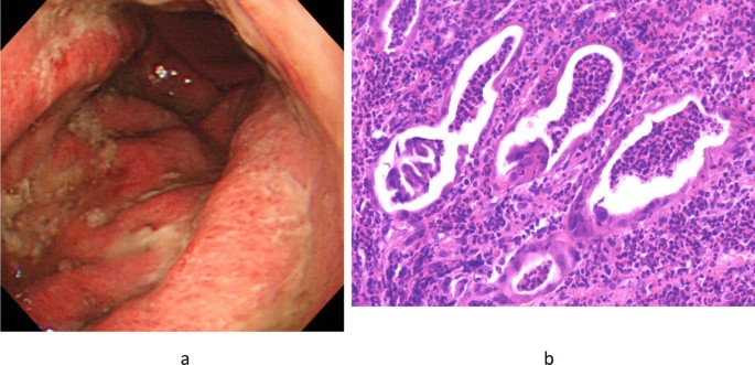
Hence, the patient underwent esophagogastroduodenoscopy, which revealed a significant hyperplasia of the gastric folds with abundant mucopus, extensive redness and edema of the gastric mucosa, and poor distensibility of the gastric wall (Fig. 1 a). Considering this endoscopic image, scirrhous gastric cancer was suspected. On histopathological examination, mucosal necrosis and severe neutrophil infiltration were detected, with no malignant cells (Fig. 1 b).

a Endoscopic image of case 1. Significant hyperplasia of the gastric folds with abundant mucopus, and extensive redness and edema of the gastric mucosa were observed. b Histological image of case 1. Mucosal necrosis and neutrophil infiltration were detected, but no malignant cells were found
The clinical course (increased fever, epigastric pain, and elevated WBC count) as well as imaging (gastric wall thickness on CT and endoscopic findings) and histopathological findings led us to consider the serious infection and to diagnose the patient with phlegmonous gastritis, despite the biopsy specimen culture being negative.
Antibiotic therapy (levofloxacin, 500 mg/day × 7 days) was then provided, leading to the patient’s recovery.
A 33-year-old Asian man with epigastralgia visited our hospital. He had a history of excessive alcohol consumption (the exact amount was unknown), had no medical history, and was sexually active. The WBC count was 12,400/mm 3 . Abdominal CT detected localized thickening of the gastric wall, mainly in the antrum.
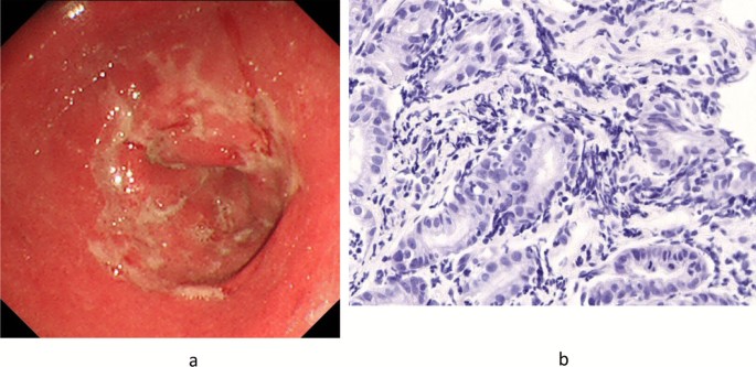
The endoscopic image revealed multiple shallow ulcers with slough fused into a map-like pattern in the same area (Fig. 2 a).

a Endoscopic image of case 2. Multiple shallow ulcers with slough fused into a map-like pattern were observed in the antrum. b Histological image of case 2. Immunostaining of the biopsy specimen was negative ( Treponema pallidum was not found)
Given the endoscopic findings, the patient was diagnosed with gastric syphilis. However, his serum test result for syphilis was negative, and the culture of biopsy specimen revealed the presence of Streptococcus viridans , resulting in the diagnosis of phlegmonous gastritis. In addition, immunostaining of the biopsy specimen was negative ( Treponema pallidum was not found) (Fig. 2 b). Thus, antibiotic therapy (amoxicillin, 750 mg/day × 14 days) was started. After 2 weeks, the endoscopic image revealed a remarkable improvement.
A 19-year-old Asian male college student visited our hospital, complaining of epigastralgia and vomiting. He had no history of drug and alcohol consumption and had no medical history. However, he had a fever (38.0 °C), with a WBC count of 15,900/mm 3 . Abdominal CT detected a thickened gastric wall, particularly in the antrum.
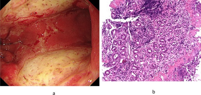
Endoscopic image showed large and shallow ulcers with reddish and edematous mucosa in the antrum, with some coagulation (Fig. 3 a). We initially suspected acute gastric mucosal lesion (AGML). However, the patient was eventually diagnosed with phlegmonous gastritis because Pseudomonas aeruginosa and S. viridans were found in the culture of gastric juice.

a Endoscopic image of case 3. Large and shallow ulcers with reddish and edematous mucosa were observed in the antrum, with some coagulation. b Histological image of case 3. Direct microscopy found no Helicobacter pylori
Meanwhile, the culture of gastric tissue was negative for Helicobacter pylori . Antibiotic therapy (tazobactam/piperacillin, 9.0 g/day × 10 days) was then started, and gastric lesions gradually disappeared.
H. pylori were not identified using direct microscopy in all cases (Fig. 3 b).
Phlegmonous gastritis is a rare inflammatory gastric disease caused by local or diffuse inflammation of the gastric wall [ 1 ]. The diffuse type is more common and has a higher mortality rate than the localized type. Case 1 was a diffuse type, whereas cases 2 and 3 showed a localized type. Imaging examinations are useful for diagnosing phlegmonous gastritis, which is suspected when gastric wall thickness is detected on abdominal ultrasonography or CT. Endoscopic ultrasonography for phlegmonous gastritis diagnosis is reportedly effective but is performed in few facilities [ 2 ].
By cause, phlegmonous gastritis is categorized into primary, secondary, and idiopathic types [ 3 , 4 ]. The most common is the primary type, which is caused by a mucosal injury such as peptic ulcer or gastric cancer. The secondary type is associated with the infection of other organs, such as biliary infection or hepatic abscess. It may also occur after endoscopic submucosal dissection and endoscopic ultrasound-guided fine-needle aspiration [ 5 , 6 ]. In the idiopathic type, as the name implies, the cause is unknown, and it mostly occurs in a compromised host. Moreover, cases 1 and 2 had a history of excessive alcohol consumption. Such a history is a causative factor of superficial gastritis, multiple small ulcers, and hemorrhagic erosions because alcohol can cause gastric mucosal injury, which induces phlegmonous gastritis [ 7 ]. Case 3 had no apparent background. However, he had an upcoming examination for promotion and felt extremely stressed. Mucosal injury such as AGML caused by mental stress could be the cause of phlegmonous gastritis [ 8 ]. Furthermore, temporary immunosuppression due to disruption in the rhythm of life might have triggered the onset of the disease in these three cases.
By clinical course, phlegmonous gastritis is categorized into acute, chronic, and subacute types [ 9 ]. All of our patients had a sudden onset of the disease, complaining of epigastralgia upon consultation. Therefore, they were categorized as the acute type.
The most common pathogenic cause of phlegmonous gastritis is Streptococcus (approximately 70%), which is resistant to gastric acid, followed by Enterococcus and Staphylococcus [ 10 ].
If the pathogen is found in the culture of gastric tissue or fluid, the diagnosis of phlegmonous gastritis could be made. However, pathogen is not always detected.
In case 1, although a pathogen was not identified, the patient was still diagnosed with phlegmonous gastritis because of other factors, such as the clinical course, symptoms, and imaging examinations.
Typically, the endoscopic image features of phlegmonous gastritis include reddish and edematous mucosa, swollen gastric folds, erosion, ulcer with slough, and mucopus adhesion. Considering that these features are nonspecific, various endoscopic images can be found in phlegmonous gastritis. Hence, we need to consider the differential diagnosis of phlegmonous gastritis.
Case 1 Scirrhous gastric cancer was suspected according to the following endoscopic image features: extensively reddish and edematous gastric mucosa, swollen gastric folds, abundant mucopus adhesion, and poor gastric wall distensibility.
Histopathologically, no malignancy was noted.
Obtaining a clinical history for several months is important because scirrhous gastric cancer takes a long time course.
Ultimately, imaging and histopathological examinations led to the diagnosis of phlegmonous gastritis.
Case 2 Gastric syphilis was suspected according to an endoscopic image feature, that is, multiple shallow ulcers with slough fused into a map-like pattern in the antrum. Furthermore, the patient was sexually active toward many unknown individuals. Regarding syphilis, the serum test and the immunostaining of the biopsy specimen were both negative ( Treponema pallidum was not found). However, S. viridans was detected in the culture; thus, the patient was diagnosed with phlegmonous gastritis.
When gastric syphilis is suspected, serum test and skin examination must be conducted.
Case 3 AGML was suspected because of the following endoscopic images: large and shallow ulcers with reddish and edematous mucosa and some signs of coagulation.
P. aeruginosa and S. viridans were found in the culture of gastric juice.
AGML is frequently caused by various factors, such as drugs (for example antibiotics, steroids, and nonsteroidal antiinflammatory agents), extreme mental or physical stress, and excessive alcohol consumption. Acute H. pylori infection must also be considered [ 11 ]. The patient had no history drug and alcohol intake and was negative for H. pylori infection. Considering that he had a history of mental stress, AGML caused by this stress was considered to be the cause of phlegmonous gastritis.
The treatment for phlegmonous gastritis is conservative treatment using antibiotics. However, surgical treatment is required in resistant or severe cases [ 12 ]. Given that fatal cases were occasionally reported, physicians need to make a correct diagnosis and start the appropriate treatment [ 13 ].
Availability of data and materials
There are no additional data available for this study.
Abbreviations
White blood cell
Computed tomography
- Acute gastric mucosal lesion
Aviles JF, Fernandez-Seara J, Barcena R, et al . Localized phlegmonous gastritis: endoscopic view. Endoscopy. 1988;20:38–9.
Article CAS Google Scholar
Hu DC, McGrath KM, Jowell PS, et al . Phlegmonous gastritis: successful treatment with antibiotics and resolution documented by EUS. Gastrointest Endosc. 2000;52:793–5.
Konjetzny GE, des Magens E. Handbuch der Speziellen Pathologischen Anatomie und Histologie, vol. 4. Berlin: Julius Springer Verlag; 1928. p. 768–1116.
Google Scholar
Ishioka M, Watanabe N, Sawaguchi M, et al . Phlegmonous gastritis: a report of three cases with clinical and imaging features. Intern Med. 2018;57:2185–8.
Article Google Scholar
Ajibe H, Osawa H, Yoshizawa M, et al . Phlegmonous gastritis after endoscopic submucosal dissection for early gastric cancer. Therap Adv Gastroentrol. 2008;1:91–5.
Itonaga M, Ueda K, Ichinose M. Phlegmonous gastritis caused by endoscopic ultrasound-guided fine-needle aspiration (EUS-FNA). Dig Endosc. 2012;24:488.
Kim GY, Ward J, Henessey B, et al . Phlegmonous gastritis: case report and review. Gastrointest Endosc. 2005;61:168–74.
Mitchell JS. Stress-related mucosal disease: risk factors and prophylactic therapy. Clin Ther. 2004;26:197–213.
Sakata T, Narita M, Ohtani N, et al . A case of phlegmonous gastritis with multiple liver and splenic abscesses. Nihon Shokakibyo Gakkai Zasshi. 2011;108:50–8.
PubMed Google Scholar
Rada-Palomino A, Muñoz-Duyos A, Pérez-Romero N, et al . Phlegmonous gastritis: a rare entity as a differential diagnostic of an acute abdomen. Description of a case and a bibliographic review. Rev Esp Enferm Dig. 2014;106:418–24.
Kamada T, Hata J, Manabe N, et al . Can dental treatment be the infection route of H. pylori transmission in adults? Three cases of acute gastric mucosal lesions after dental treatment. Dig Endosc. 2007;19:32–5.
Yokota T, Yamaki T, Yashima R, et al . Emergency operation for phlegmonous gastritis. Ups J Med Sci. 2005;110:237–40.
Morimoto M, Tamura S, Hayakawa T, et al . Phlegmonous gastritis associated with group a streptococcal toxic shock syndrome. Intern Med. 2014;53:2639–42.
Download references
Acknowledgements
We would like to thank Dr. Tomoaki Yamasaki of Osaka City General Hospital for correcting the English used in this manuscript.
No funding.
Author information
Authors and affiliations.
Department of Internal Medicine, Higashi-Osaka Hospital, 7-22, Chuo 1 chome, Joto-ku, Osaka, 536-0005, Japan
Yoshikazu Yakami, Toshihiko Yagyu & Tomoki Bando
You can also search for this author in PubMed Google Scholar
Contributions
YY was a major contributor in writing the manuscript. TY and TB made appropriate suggestions during the preparation of the manuscript. All authors read and approved the final manuscript.
Corresponding author
Correspondence to Yoshikazu Yakami .
Ethics declarations
Ethics approval and consent to participate.
Ethical approval was not relevant or applicable to this case report.
Consent for publication
Written informed consent was obtained from the patients for publication of this case report and any accompanying images. A copy of the written consent is available for review by the Editor-in-Chief of this journal.
Competing interests
The authors declare that they have no competing interests.
Additional information
Publisher’s note.
Springer Nature remains neutral with regard to jurisdictional claims in published maps and institutional affiliations.

Rights and permissions
Open Access This article is licensed under a Creative Commons Attribution 4.0 International License, which permits use, sharing, adaptation, distribution and reproduction in any medium or format, as long as you give appropriate credit to the original author(s) and the source, provide a link to the Creative Commons licence, and indicate if changes were made. The images or other third party material in this article are included in the article's Creative Commons licence, unless indicated otherwise in a credit line to the material. If material is not included in the article's Creative Commons licence and your intended use is not permitted by statutory regulation or exceeds the permitted use, you will need to obtain permission directly from the copyright holder. To view a copy of this licence, visit http://creativecommons.org/licenses/by/4.0/ . The Creative Commons Public Domain Dedication waiver ( http://creativecommons.org/publicdomain/zero/1.0/ ) applies to the data made available in this article, unless otherwise stated in a credit line to the data.
Reprints and permissions
About this article
Cite this article.
Yakami, Y., Yagyu, T. & Bando, T. Phlegmonous gastritis: a case series. J Med Case Reports 15 , 445 (2021). https://doi.org/10.1186/s13256-021-02999-9
Download citation
Received : 13 July 2020
Accepted : 05 July 2021
Published : 06 September 2021
DOI : https://doi.org/10.1186/s13256-021-02999-9
Share this article
Anyone you share the following link with will be able to read this content:
Sorry, a shareable link is not currently available for this article.
Provided by the Springer Nature SharedIt content-sharing initiative
- Phlegmonous gastritis
- Scirrhous gastric cancer
- Gastric syphilis
Journal of Medical Case Reports
ISSN: 1752-1947
- Submission enquiries: Access here and click Contact Us
- General enquiries: [email protected]
An official website of the United States government
The .gov means it’s official. Federal government websites often end in .gov or .mil. Before sharing sensitive information, make sure you’re on a federal government site.
The site is secure. The https:// ensures that you are connecting to the official website and that any information you provide is encrypted and transmitted securely.
- Publications
- Account settings
Preview improvements coming to the PMC website in October 2024. Learn More or Try it out now .
- Advanced Search
- Journal List
- v.30(7); 1989 Jul

Chronic gastritis and gastroduodenal ulcer: a case control study on risk of coexisting duodenal or gastric ulcer in patients with gastritis.
Chronic (atrophic) gastritis (AG) is common in active duodenal (DU) and gastric ulcer (GU) disease. In this case control study in consecutive prospective outpatients (571 cases and 1074 controls) who had undergone diagnostic upper gastrointestinal endoscopy and routine biopsies from both antral and body mucosa, we calculated the risk of coexisting active DU and/or GU in different gastritis of the antrum or body and according to grade (superficial gastritis, mild, moderate or severe atrophic gastritis). The risk of coexisting active gastroduodenal ulcer (ulcer in duodenum and/or stomach), as well as the risk of DU or GU, was dependent upon the presence and grade of gastritis in antrum and body mucosa. The risk of coexisting ulcer, as expressed as an age adjusted relative risk (RR) and calculated as odds ratio of gastritis in cases and controls, was significantly increased in the presence of superficial antral and body gastritis (RR = 8.5 (7.0-20.0) in men; RR = 5.8 (3.3-10.2) in women), as compared with the risk of ulcer in subjects with histologically normal mucosa (RR = 1). The risk of ulcer, and the risk of GU in particular, increased further with increasing severity of antral gastritis. In such patients with moderate or severe atrophic antral gastritis the RR of coexisting ulcer even exceeded 20 in men and 10 in women (RR = 25.6 (9.0-72.7) in men; RR = 11.7 (5.9-23.0) in women). On the other hand, the RR of ulcer, and the RR of DU in particular, was below 1 in the presence of atrophic gastritis in the gastric body, irrespective of the grade of gastritis in the antrum. We conclude that the type and grade of gastritis strongly predicts the risk of coexisting peptic ulcer, and that the risk of coexisting DU or GU increases with an increase in grade of AG of the antrum but decreases with an increase in grade of AG of the gastric body.
Full text is available as a scanned copy of the original print version. Get a printable copy (PDF file) of the complete article (1.0M), or click on a page image below to browse page by page. Links to PubMed are also available for Selected References .
Selected References
These references are in PubMed. This may not be the complete list of references from this article.
- Mackay IR, Hislop IG. Chronic gastritis and gastric ulcer. Gut. 1966 Jun; 7 (3):228–233. [ PMC free article ] [ PubMed ] [ Google Scholar ]
- Schrager J, Spink R, Mitra S. The antrum in patients with duodenal and gastric ulcers. Gut. 1967 Oct; 8 (5):497–508. [ PMC free article ] [ PubMed ] [ Google Scholar ]
- Gear MW, Truelove SC, Whitehead R. Gastric ulcer and gastritis. Gut. 1971 Aug; 12 (8):639–645. [ PMC free article ] [ PubMed ] [ Google Scholar ]
- Meister H, Holubarsch C, Haferkamp O, Schlag P, Herfarth C. Gastritis, intestinal metaplasia and dysplasia versus benign ulcer in stomach and duodenum and gastric carcinoma -- a histotopographical study. Pathol Res Pract. 1979 Jan; 164 (3):259–269. [ PubMed ] [ Google Scholar ]
- Nielsen HO, Muñoz JD, Kronborg O, Andersen D. The antrum in duodenal ulcer patients. Relationship between antrum size, nerve of Latarjet, gastrin cell quantity, and gastric acid secretion. Scand J Gastroenterol. 1981; 16 (4):491–495. [ PubMed ] [ Google Scholar ]
- Earlam RJ, Amerigo J, Kakavoulis T, Pollock DJ. Histological appearances of oesophagus, antrum and duodenum and their correlation with symptoms in patients with a duodenal ulcer. Gut. 1985 Jan; 26 (1):95–100. [ PMC free article ] [ PubMed ] [ Google Scholar ]
- Meikle DD, Taylor KB, Truelove SC, Whitehead R. Gastritis duodenitis, and circulating levels of gastrin in duodenal ulcer before and after vagotomy. Gut. 1976 Sep; 17 (9):719–728. [ PMC free article ] [ PubMed ] [ Google Scholar ]
- Cheli R, Giacosa A. Duodenal ulcer and chronic gastritis. Endoscopy. 1986 Jul; 18 (4):125–126. [ PubMed ] [ Google Scholar ]
- Hui WM, Lam SK, Ho J, Ng MM, Lui I, Lai CL, Lok AS, Lau WY, Poon GP, Choi S, et al. Chronic antral gastritis in duodenal ulcer. Natural history and treatment with prostaglandin E1. Gastroenterology. 1986 Nov; 91 (5):1095–1101. [ PubMed ] [ Google Scholar ]
- Ippoliti A, Walsh J. Newer concepts in the pathogenesis of peptic ulcer disease. Surg Clin North Am. 1976 Dec; 56 (6):1479–1490. [ PubMed ] [ Google Scholar ]
- Varis K, Ihamäki T, Härkönen M, Samloff IM, Siurala M. Gastric morphology, function, and immunology in first-degree relatives of probands with pernicious anemia and controls. Scand J Gastroenterol. 1979; 14 (2):129–139. [ PubMed ] [ Google Scholar ]
- Siurala M, Isokoski M, Varis K, Kekki M. Prevalence of gastritis in a rural population. Bioptic study of subjects selected at random. Scand J Gastroenterol. 1968; 3 (2):211–223. [ PubMed ] [ Google Scholar ]
- Whitehead R, Truelove SC, Gear MW. The histological diagnosis of chronic gastritis in fibreoptic gastroscope biopsy specimens. J Clin Pathol. 1972 Jan; 25 (1):1–11. [ PMC free article ] [ PubMed ] [ Google Scholar ]
- Hovinen E, Kekki M, Kuikka S. A theory to the stochastic dynamic model building for chronic progressive disease processes with an application to chronic gastritis. J Theor Biol. 1976 Mar; 57 (1):131–152. [ PubMed ] [ Google Scholar ]
- Siurala M, Varis K, Kekki M. New aspects on epidemiology, genetics, and dynamics of chronic gastritis. Front Gastrointest Res. 1980; 6 :148–166. [ PubMed ] [ Google Scholar ]
- Kekki M, Sipponen P, Siurala M. Progression of antral and body gastritis in patients with active and healed duodenal ulcer and duodenitis. Scand J Gastroenterol. 1984 May; 19 (3):382–388. [ PubMed ] [ Google Scholar ]
- Strickland RG, Mackay IR. A reappraisal of the nature and significance of chronic atrophic gastritis. Am J Dig Dis. 1973 May; 18 (5):426–440. [ PubMed ] [ Google Scholar ]
- Moore SC, Malagelada JR, Shorter RG, Zinsmeister AR. Interrelationships among gastric mucosal morphology, secretion, and motility in peptic ulcer disease. Dig Dis Sci. 1986 Jul; 31 (7):673–684. [ PubMed ] [ Google Scholar ]
- Piper DW, Greig M, Coupland GA, Hobbin E, Shinners J. Factors relevant to the prognosis of chronic gastric ulcer. Gut. 1975 Sep; 16 (9):714–718. [ PMC free article ] [ PubMed ] [ Google Scholar ]
- Maaroos HI, Salupere V, Uibo R, Kekki M, Sipponen P. Seven-year follow-up study of chronic gastritis in gastric ulcer patients. Scand J Gastroenterol. 1985 Mar; 20 (2):198–204. [ PubMed ] [ Google Scholar ]
- Oi M, Ito Y, Kumagai F, Yoshida K, Tanaka Y, Yoshikawa K, Miho O, Kijima M. A possible dual control mechanism in the origin of peptic ulcer. A study on ulcer location as affected by mucosa and musculature. Gastroenterology. 1969 Sep; 57 (3):280–293. [ PubMed ] [ Google Scholar ]
- Stemmermann G, Haenszel W, Locke F. Epidemiologic pathology of gastric ulcer and gastric carcinoma among Japanese in Hawaii. J Natl Cancer Inst. 1977 Jan; 58 (1):13–20. [ PubMed ] [ Google Scholar ]
- Kimura K. Chronological transition of the fundic-pyloric border determined by stepwise biopsy of the lesser and greater curvatures of the stomach. Gastroenterology. 1972 Oct; 63 (4):584–592. [ PubMed ] [ Google Scholar ]
- Trier JS. Morphology of the gastric mucosa in patients with ulcer diseases. Am J Dig Dis. 1976 Feb; 21 (2):138–140. [ PubMed ] [ Google Scholar ]
- Stadelmann O, Elster K, Stolte M, Miederer SE, Deyhle P, Demling L, Siegenthaler W. The peptic gastric ulcer--histotopographic and functional investigations. Scand J Gastroenterol. 1971; 6 (7):613–623. [ PubMed ] [ Google Scholar ]
- Cheng FC, Lam SK, Ong GB. Maximum acid output to graded doses of pentagastrin and its relation to parietal cell mass in Chinese patients with duodenal ulcer. Gut. 1977 Oct; 18 (10):827–832. [ PMC free article ] [ PubMed ] [ Google Scholar ]
- CARD WI, MARKS IN. The relationship between the acid output of the stomach following "maximal" histamine stimulation and the parietal cell mass. Clin Sci. 1960 Feb; 19 :147–163. [ PubMed ] [ Google Scholar ]
- Samloff IM, Stemmermann GN, Heilbrun LK, Nomura A. Elevated serum pepsinogen I and II levels differ as risk factors for duodenal ulcer and gastric ulcer. Gastroenterology. 1986 Mar; 90 (3):570–576. [ PubMed ] [ Google Scholar ]
Ohio State nav bar
The Ohio State University
- BuckeyeLink
- Find People
- Search Ohio State
Patient Case Presentation
Vital Signs and Measurements
- BP: 135/85 mm Hg
- HR: 99 bpm
- RR: 21 b / min
- Temperature: 36.1 ℃
- Pulse oxygenation: 99%
- Height: 167 cm
- Weight: 58.2 kg
Past medical history
- Gastritis with Helicobacter pylori (H. pylori) infection, diagnosed 5 years ago. Resolved with pharmacotherapy, frequent recurrence.
- Heart attack 3 months ago, has started taking aspirin since then.
- Osteoarthritis, diagnosed 3 years ago. Long-term use of the non-steroidal anti-inflammatory drug (NSAID) since diagnosis.
- Acute pancreatitis 5 years ago, resolved with pharmacotherapy, no recurrence.
- Chronic obstructive pulmonary disease, diagnosed 11 years ago.
- Diabetes Mellitus and hypertension diagnosed 6 years ago.
- No surgical history.
Pertinent family history
- Mother died from gastric cancer at age 67 years old.
- Father alive, has smoked for 50 years and now has a small-cell carcinoma. Live with the patient since she divorced her husband.
- Brother alive and well at age 56 years, had a history of duodenal ulcers. Heavy smoker.
- Ex-husband at age 62, alive and has COPD.
- One son alive and well at age 29.
- One daughter at age 35, alive and has recurrent Gastritis.
Pertinent social history
- Has worked full-time as a TV program producer for 30 years.
- Divorced with her husband 20 years prior, raised 2 children by herself.
- Hobbies include drinking, eating spicy food, watching dramas and talkshow.
- Has smoked for 30 years, even after the diagnosis of COPD, still cannot quit smoking.
A Patient/Family Care Study on Gastritis
Original bundle.
License bundle
Collections.
Academia.edu no longer supports Internet Explorer.
To browse Academia.edu and the wider internet faster and more securely, please take a few seconds to upgrade your browser .
Enter the email address you signed up with and we'll email you a reset link.
- We're Hiring!
- Help Center

Case Study on Acute Gastroenteritis

Gastroenteritis, sometimes referred to as “stomach flu”, is an inflammation of the GI (gastrointestinal) tract, which includes the stomach and intestines. Most cases of gastroenteritis are caused by viruses. Bacterial gastroenteritis (caused by bacteria) often causes severe symptoms. It can even be fatal. It is also the most common digestive disorder among children. Severe gastroenteritis causes dehydration and an imbalance of blood chemicals (electrolytes) because of a loss of body fluids in the vomit and stool. This can be acquired through contaminated food and water that contains harmful bacteria (such as salmonella, Campylobacter, and E. coli). Food can be contaminated when food handlers don’t wash their hands. Or when food isn’t stored, handled, or cooked correctly. This can also be acquired and spread through the fecal-oral route, people with gastroenteritis have harmful bacteria in their stool. When they don’t wash their hands well after using the bathroom, they can spread the germs to objects. If you touch the same objects, you can pick up the germs on your hands and transfer them to your mouth. (Fairview.org, 2021)
RELATED PAPERS
Physical Review E
Edgar Manuel Cahun Martin
Proceedings of the 9th IEEE International Conference on Collaborative Computing: Networking, Applications and Worksharing
Sandro Nielsen
Monographien
Stefan Schleicher
Revista HISTEDBR On-line
Marcela Rebeca Contreras Loera
PRODUSEN RAK BUNGA ALUMINIUM
Family & Community Health
Courtney FitzGerald
Journal of gastroenterology and hepatology research
Mohamed Elwany
Seminario De Pesquisa Em Estudos Linguisticos
IVETE SANTOS
Strojarstvo: časopis za …
Sandra Milunović
SN Computer Science
Muhammad Sadiq jibril
Seminary Library
Hospital topics
alberto coustasse
Taufiq rahman
International Journal of Computer Applications
Rishit Jain
arXiv (Cornell University)
Fei Victor LIM
Journal of The Saudi Heart Association
Akram Fayed
Danilo Marcondes Filho
Ingrid Scheurmann
WSEAS transactions on advances in engineering education
MD.Sagor Mahmud
Sigit Priyanto
44th Biennial Convention (28 October - 01 November 2017)
Wyona M Freysteinson, PhD, MN
BioPsychoSocial Medicine
Hamid Afshar
Jules Vivid
See More Documents Like This
RELATED TOPICS
- We're Hiring!
- Help Center
- Find new research papers in:
- Health Sciences
- Earth Sciences
- Cognitive Science
- Mathematics
- Computer Science
- Academia ©2024

IMAGES
VIDEO
COMMENTS
case study gastritis - Free download as Powerpoint Presentation (.ppt / .pptx), PDF File (.pdf), Text File (.txt) or view presentation slides online. Scribd is the world's largest social reading and publishing site.
Case Study of Acute Gastritis - Free download as Word Doc (.doc / .docx), PDF File (.pdf), Text File (.txt) or read online for free. for educational purposes only
Background Phlegmonous gastritis is a rare and fatal infectious disease of the stomach, presenting varied and nonspecific endoscopic images, which are therefore difficult to diagnose. This report discusses three cases of phlegmonous gastritis, each with unique endoscopic images, and considers the differential diagnosis of this disease. These cases were initially suspected of scirrhous gastric ...
1. INTRODUCTION. Acute phlegmonous gastritis (APG) is a rare and often fatal disease. Symptoms can be non‐specific. Patients typically present with abdominal pain, nausea, vomiting, fever, and signs of infection. 1 The most common pathogens related to APG described in the literature are the Streptococcus species. 1 , 3 Although the ...
Acute gastritis is a term that encompasses a broad spectrum of entities that induce inflammatory changes in the gastric mucosa. Several different etiologies share the same general clinical presentation; however, they differ in their unique histologic characteristics. The inflammation may involve the entire stomach (eg, pangastritis) or a region ...
The pain woke her from sleep and was 10/10. She was taken to her local hospital's emergency department, where an abdominal computed tomography (CT) scan showed no radiographic evidence of appendicitis but did show some gastric thickening. Laboratory studies showed an elevated white blood cell count (18.0) and an increased sedimentation rate.
Physical examination. The physical examination findings are often normal, with occasional mild epigastric tenderness. The examination tends to exhibit more abnormalities as the patient develops complications in relation to gastritis. Differential Diagnoses. Acute gastritis is a term covering a broad spectrum of entities that induce inflammatory ...
Abstract. Chronic (atrophic) gastritis (AG) is common in active duodenal (DU) and gastric ulcer (GU) disease. In this case control study in consecutive prospective outpatients (571 cases and 1074 controls) who had undergone diagnostic upper gastrointestinal endoscopy and routine biopsies from both antral and body mucosa, we calculated the risk of coexisting active DU and/or GU in different ...
Assessment and Interventions: Obtain the patient's weight and other body measurements. The admission weight serves as a baseline metric and helps guide interventions. Obtain information about the patient's eating habits. Some food items can exacerbate the symptoms of gastritis. Acidic or citrus food items may worsen symptoms.
Scribd adalah situs bacaan dan penerbitan sosial terbesar di dunia.
GI case study questions. Peptic ulcer- caused by h pylori or gastritis. NSAIDS are also a cause - second leading. h pylori is a gram negative bacteria -is very common. H Pylori- key culprit. Bacteria eats away at lining of the stomach or duodenum.
Patient Case Presentation. A 61-year-old American woman was referred to a Gastroenterology Clinic from primary care provider due to consistent discomfort and significant weight loss. She looked for a PCP's advice as she had a tarry stool in the early morning which she had never experienced before. She presented with a 2-month history of ...
The study provides a systematic way of collecting data, analysing information, and reporting the results of nursing care. This Patient/Family care study is based on holistic care, taking into account all factors impinging on the health of the patient. The Patient/Family care study forms an integral part of the curriculum for educating nursing ...
Gastroenteritis, sometimes referred to as "stomach flu", is an inflammation of the GI (gastrointestinal) tract, which includes the stomach and intestines. Most cases of gastroenteritis are caused by viruses. Bacterial gastroenteritis (caused by bacteria) often causes severe symptoms. It can even be fatal.
Diagnosis. Gastritis is defined as inflammation of the stomach, where Helicobacter pylori (H. pylori) is the most common cause of Gastritis. Gastritis could be sudden ( acute ), but it could also appear slowly over time ( chronic ). With unproper treatment or not treating gastritis, patients with gastritis could experience exacerbation.