Introduction to the Nervous System
The nervous system is the major controlling, regulatory, and communicating system in the body. It is the center of all mental activity including thought, learning, and memory. Together with the endocrine system , the nervous system is responsible for regulating and maintaining homeostasis . Through its receptors, the nervous system keeps us in touch with our environment, both external and internal .
Like other systems in the body, the nervous system is composed of organs, principally the brain , spinal cord , nerves, and ganglia . These, in turn, consist of various tissues, including nerve , blood , and connective tissue . Together these carry out the complex activities of the nervous system.
The various activities of the nervous system can be grouped together as three general, overlapping functions:
- Integrative
Millions of sensory receptors detect changes, called stimuli, which occur inside and outside the body. They monitor such things as temperature, light, and sound from the external environment. Inside the body, the internal environment, receptors detect variations in pressure, pH , carbon dioxide concentration, and the levels of various electrolytes . All of this gathered information is called sensory input.
Sensory input is converted into electrical signals called nerve impulses that are transmitted to the brain. There the signals are brought together to create sensations, to produce thoughts, or to add to memory; Decisions are made each moment based on the sensory input. This is integration.
Based on the sensory input and integration, the nervous system responds by sending signals to muscles, causing them to contract, or to glands, causing them to produce secretions. Muscles and glands are called effectors because they cause an effect in response to directions from the nervous system. This is the motor output or motor function.

12.1 Basic Structure and Function of the Nervous System
Learning objectives.
By the end of this section, you will be able to:
- Identify the anatomical and functional divisions of the nervous system
- Relate the functional and structural differences between gray matter and white matter structures of the nervous system to the structure of neurons
- List the basic functions of the nervous system
The picture you have in your mind of the nervous system probably includes the brain , the nervous tissue contained within the cranium, and the spinal cord , the extension of nervous tissue within the vertebral column. That suggests it is made of two organs—and you may not even think of the spinal cord as an organ—but the nervous system is a very complex structure. Within the brain, many different and separate regions are responsible for many different and separate functions. It is as if the nervous system is composed of many organs that all look similar and can only be differentiated using tools such as the microscope or electrophysiology. In comparison, it is easy to see that the stomach is different than the esophagus or the liver, so you can imagine the digestive system as a collection of specific organs.
The Central and Peripheral Nervous Systems
The nervous system can be divided into two major regions: the central and peripheral nervous systems. The central nervous system (CNS) is the brain and spinal cord, and the peripheral nervous system (PNS) is everything else ( Figure 12.2 ). The brain is contained within the cranial cavity of the skull, and the spinal cord is contained within the vertebral cavity of the vertebral column. It is a bit of an oversimplification to say that the CNS is what is inside these two cavities and the peripheral nervous system is outside of them, but that is one way to start to think about it. In actuality, there are some elements of the peripheral nervous system that are within the cranial or vertebral cavities. The peripheral nervous system is so named because it is on the periphery—meaning beyond the brain and spinal cord. Depending on different aspects of the nervous system, the dividing line between central and peripheral is not necessarily universal.
Nervous tissue, present in both the CNS and PNS, contains two basic types of cells: neurons and glial cells. A glial cell is one of a variety of cells that provide a framework of tissue that supports the neurons and their activities. The neuron is the more functionally important of the two, in terms of the communicative function of the nervous system. To describe the functional divisions of the nervous system, it is important to understand the structure of a neuron. Neurons are cells and therefore have a soma , or cell body, but they also have extensions of the cell; each extension is generally referred to as a process . There is one important process that every neuron has called an axon , which is the fiber that connects a neuron with its target. Another type of process that branches off from the soma is the dendrite . Dendrites are responsible for receiving most of the input from other neurons. Looking at nervous tissue, there are regions that predominantly contain cell bodies and regions that are largely composed of just axons. These two regions within nervous system structures are often referred to as gray matter (the regions with many cell bodies and dendrites) or white matter (the regions with many axons). Figure 12.3 demonstrates the appearance of these regions in the brain and spinal cord. The colors ascribed to these regions are what would be seen in “fresh,” or unstained, nervous tissue. Gray matter is not necessarily gray. It can be pinkish because of blood content, or even slightly tan, depending on how long the tissue has been preserved. But white matter is white because axons are insulated by a lipid-rich substance called myelin . Lipids can appear as white (“fatty”) material, much like the fat on a raw piece of chicken or beef. Actually, gray matter may have that color ascribed to it because next to the white matter, it is just darker—hence, gray.
The distinction between gray matter and white matter is most often applied to central nervous tissue, which has large regions that can be seen with the unaided eye. When looking at peripheral structures, often a microscope is used and the tissue is stained with artificial colors. That is not to say that central nervous tissue cannot be stained and viewed under a microscope, but unstained tissue is most likely from the CNS—for example, a frontal section of the brain or cross section of the spinal cord.
Regardless of the appearance of stained or unstained tissue, the cell bodies of neurons or axons can be located in discrete anatomical structures that need to be named. Those names are specific to whether the structure is central or peripheral. A localized collection of neuron cell bodies in the CNS is referred to as a nucleus . In the PNS, a cluster of neuron cell bodies is referred to as a ganglion . Figure 12.4 indicates how the term nucleus has a few different meanings within anatomy and physiology. It is the center of an atom, where protons and neutrons are found; it is the center of a cell, where the DNA is found; and it is a center of some function in the CNS. There is also a potentially confusing use of the word ganglion (plural = ganglia) that has a historical explanation. In the central nervous system, there is a group of nuclei that are connected together and were once called the basal ganglia before “ganglion” became accepted as a description for a peripheral structure. Some sources refer to this group of nuclei as the “basal nuclei” to avoid confusion.
Terminology applied to bundles of axons also differs depending on location. A bundle of axons, or fibers, found in the CNS is called a tract whereas the same thing in the PNS would be called a nerve . There is an important point to make about these terms, which is that they can both be used to refer to the same bundle of axons. When those axons are in the PNS, the term is nerve, but if they are CNS, the term is tract. The most obvious example of this is the axons that project from the retina into the brain. Those axons are called the optic nerve as they leave the eye, but when they are inside the cranium, they are referred to as the optic tract. There is a specific place where the name changes, which is the optic chiasm, but they are still the same axons ( Figure 12.5 ). A similar situation outside of science can be described for some roads. Imagine a road called “Broad Street” in a town called “Anyville.” The road leaves Anyville and goes to the next town over, called “Hometown.” When the road crosses the line between the two towns and is in Hometown, its name changes to “Main Street.” That is the idea behind the naming of the retinal axons. In the PNS, they are called the optic nerve, and in the CNS, they are the optic tract. Table 12.1 helps to clarify which of these terms apply to the central or peripheral nervous systems.
Interactive Link
In 2003, the Nobel Prize in Physiology or Medicine was awarded to Paul C. Lauterbur and Sir Peter Mansfield for discoveries related to magnetic resonance imaging (MRI). This is a tool to see the structures of the body (not just the nervous system) that depends on magnetic fields associated with certain atomic nuclei. The utility of this technique in the nervous system is that fat tissue and water appear as different shades between black and white. Because white matter is fatty (from myelin) and gray matter is not, they can be easily distinguished in MRI images. Try this PhET simulation that demonstrates the use of this technology and compares it with other types of imaging technologies. Also, the results from an MRI session are compared with images obtained from X-ray or computed tomography. How do the imaging techniques shown in this game indicate the separation of white and gray matter compared with the freshly dissected tissue shown earlier?
Functional Divisions of the Nervous System
The nervous system can also be divided on the basis of its functions, but anatomical divisions and functional divisions are different. The CNS and the PNS both contribute to the same functions, but those functions can be attributed to different regions of the brain (such as the cerebral cortex or the hypothalamus) or to different ganglia in the periphery. The problem with trying to fit functional differences into anatomical divisions is that sometimes the same structure can be part of several functions. For example, the optic nerve carries signals from the retina that are either used for the conscious perception of visual stimuli, which takes place in the cerebral cortex, or for the reflexive responses of smooth muscle tissue that are processed through the hypothalamus.
There are two ways to consider how the nervous system is divided functionally. First, the basic functions of the nervous system are sensation, integration, and response. Secondly, control of the body can be somatic or autonomic—divisions that are largely defined by the structures that are involved in the response. There is also a region of the peripheral nervous system that is called the enteric nervous system that is responsible for a specific set of the functions within the realm of autonomic control related to gastrointestinal functions.
Basic Functions
The nervous system is involved in receiving information about the environment around us (sensation) and generating responses to that information (motor responses). The nervous system can be divided into regions that are responsible for sensation (sensory functions) and for the response (motor functions). But there is a third function that needs to be included. Sensory input needs to be integrated with other sensations, as well as with memories, emotional state, or learning (cognition). Some regions of the nervous system are termed integration or association areas. The process of integration combines sensory perceptions and higher cognitive functions such as memories, learning, and emotion to produce a response.
Sensation. The first major function of the nervous system is sensation—receiving information about the environment to gain input about what is happening outside the body (or, sometimes, within the body). The sensory functions of the nervous system register the presence of a change from homeostasis or a particular event in the environment, known as a stimulus . The senses we think of most are the “big five”: taste, smell, touch, sight, and hearing. The stimuli for taste and smell are both chemical substances (molecules, compounds, ions, etc.), touch is physical or mechanical stimuli that interact with the skin, sight is light stimuli, and hearing is the perception of sound, which is a physical stimulus similar to some aspects of touch. There are actually more senses than just those, but that list represents the major senses. Those five are all senses that receive stimuli from the outside world, and of which there is conscious perception. Additional sensory stimuli might be from the internal environment (inside the body), such as the stretch of an organ wall or the concentration of certain ions in the blood.
Response. The nervous system produces a response on the basis of the stimuli perceived by sensory structures. An obvious response would be the movement of muscles, such as withdrawing a hand from a hot stove, but there are broader uses of the term. The nervous system can cause the contraction of all three types of muscle tissue. For example, skeletal muscle contracts to move the skeleton, cardiac muscle is influenced as heart rate increases during exercise, and smooth muscle contracts as the digestive system moves food along the digestive tract. Responses also include the neural control of glands in the body as well, such as the production and secretion of sweat by the eccrine and merocrine sweat glands found in the skin to lower body temperature.
Responses can be divided into those that are voluntary or conscious (contraction of skeletal muscle) and those that are involuntary (contraction of smooth muscles, regulation of cardiac muscle, activation of glands). Voluntary responses are governed by the somatic nervous system and involuntary responses are governed by the autonomic nervous system, which are discussed in the next section.
Integration. Stimuli that are received by sensory structures are communicated to the nervous system where that information is processed. This is called integration. Stimuli are compared with, or integrated with, other stimuli, memories of previous stimuli, or the state of a person at a particular time. This leads to the specific response that will be generated. Seeing a baseball pitched to a batter will not automatically cause the batter to swing. The trajectory of the ball and its speed will need to be considered. Maybe the count is three balls and one strike, and the batter wants to let this pitch go by in the hope of getting a walk to first base. Or maybe the batter’s team is so far ahead, it would be fun to just swing away.
Controlling the Body
The nervous system can be divided into two parts mostly on the basis of a functional difference in responses. The somatic nervous system (SNS) is responsible for conscious perception and voluntary motor responses. Voluntary motor response means the contraction of skeletal muscle, but those contractions are not always voluntary in the sense that you have to want to perform them. Some somatic motor responses are reflexes, and often happen without a conscious decision to perform them. If your friend jumps out from behind a corner and yells “Boo!” you will be startled and you might scream or leap back. You didn’t decide to do that, and you may not have wanted to give your friend a reason to laugh at your expense, but it is a reflex involving skeletal muscle contractions. Other motor responses become automatic (in other words, unconscious) as a person learns motor skills (referred to as “habit learning” or “procedural memory”).
The autonomic nervous system (ANS) is responsible for involuntary control of the body, usually for the sake of homeostasis (regulation of the internal environment). Sensory input for autonomic functions can be from sensory structures tuned to external or internal environmental stimuli. The motor output extends to smooth and cardiac muscle as well as glandular tissue. The role of the autonomic system is to regulate the organ systems of the body, which usually means to control homeostasis. Sweat glands, for example, are controlled by the autonomic system. When you are hot, sweating helps cool your body down. That is a homeostatic mechanism. But when you are nervous, you might start sweating also. That is not homeostatic, it is the physiological response to an emotional state.
There is another division of the nervous system that describes functional responses. The enteric nervous system (ENS) is responsible for controlling the smooth muscle and glandular tissue in your digestive system. It is a large part of the PNS, and is not dependent on the CNS. It is sometimes valid, however, to consider the enteric system to be a part of the autonomic system because the neural structures that make up the enteric system are a component of the autonomic output that regulates digestion. There are some differences between the two, but for our purposes here there will be a good bit of overlap. See Figure 12.6 for examples of where these divisions of the nervous system can be found.
Visit this site to read about a woman that notices that her daughter is having trouble walking up the stairs. This leads to the discovery of a hereditary condition that affects the brain and spinal cord. The electromyography and MRI tests indicated deficiencies in the spinal cord and cerebellum, both of which are responsible for controlling coordinated movements. To what functional division of the nervous system would these structures belong?
Everyday Connection
How much of your brain do you use.
Have you ever heard the claim that humans only use 10 percent of their brains? Maybe you have seen an advertisement on a website saying that there is a secret to unlocking the full potential of your mind—as if there were 90 percent of your brain sitting idle, just waiting for you to use it. If you see an ad like that, don’t click. It isn’t true.
An easy way to see how much of the brain a person uses is to take measurements of brain activity while performing a task. An example of this kind of measurement is functional magnetic resonance imaging (fMRI), which generates a map of the most active areas and can be generated and presented in three dimensions ( Figure 12.7 ). This procedure is different from the standard MRI technique because it is measuring changes in the tissue in time with an experimental condition or event.
The underlying assumption is that active nervous tissue will have greater blood flow. By having the subject perform a visual task, activity all over the brain can be measured. Consider this possible experiment: the subject is told to look at a screen with a black dot in the middle (a fixation point). A photograph of a face is projected on the screen away from the center. The subject has to look at the photograph and decipher what it is. The subject has been instructed to push a button if the photograph is of someone they recognize. The photograph might be of a celebrity, so the subject would press the button, or it might be of a random person unknown to the subject, so the subject would not press the button.
In this task, visual sensory areas would be active, integrating areas would be active, motor areas responsible for moving the eyes would be active, and motor areas for pressing the button with a finger would be active. Those areas are distributed all around the brain and the fMRI images would show activity in more than just 10 percent of the brain (some evidence suggests that about 80 percent of the brain is using energy—based on blood flow to the tissue—during well-defined tasks similar to the one suggested above). This task does not even include all of the functions the brain performs. There is no language response, the body is mostly lying still in the MRI machine, and it does not consider the autonomic functions that would be ongoing in the background.
As an Amazon Associate we earn from qualifying purchases.
This book may not be used in the training of large language models or otherwise be ingested into large language models or generative AI offerings without OpenStax's permission.
Want to cite, share, or modify this book? This book uses the Creative Commons Attribution License and you must attribute OpenStax.
Access for free at https://openstax.org/books/anatomy-and-physiology-2e/pages/1-introduction
- Authors: J. Gordon Betts, Kelly A. Young, James A. Wise, Eddie Johnson, Brandon Poe, Dean H. Kruse, Oksana Korol, Jody E. Johnson, Mark Womble, Peter DeSaix
- Publisher/website: OpenStax
- Book title: Anatomy and Physiology 2e
- Publication date: Apr 20, 2022
- Location: Houston, Texas
- Book URL: https://openstax.org/books/anatomy-and-physiology-2e/pages/1-introduction
- Section URL: https://openstax.org/books/anatomy-and-physiology-2e/pages/12-1-basic-structure-and-function-of-the-nervous-system
© Dec 19, 2023 OpenStax. Textbook content produced by OpenStax is licensed under a Creative Commons Attribution License . The OpenStax name, OpenStax logo, OpenStax book covers, OpenStax CNX name, and OpenStax CNX logo are not subject to the Creative Commons license and may not be reproduced without the prior and express written consent of Rice University.
If you're seeing this message, it means we're having trouble loading external resources on our website.
If you're behind a web filter, please make sure that the domains *.kastatic.org and *.kasandbox.org are unblocked.
To log in and use all the features of Khan Academy, please enable JavaScript in your browser.
High school biology
Course: high school biology > unit 8, structure of the nervous system.
- Anatomy of a neuron
- Intro to the endocrine system
- The nervous and endocrine systems review
- The nervous and endocrine systems
Want to join the conversation?
- Upvote Button navigates to signup page
- Downvote Button navigates to signup page
- Flag Button navigates to signup page

Video transcript
12.1 Structure and Function of the Nervous System
Learning objectives.
By the end of this section, you will be able to:
Relate the anatomical structures to the basic functions of the nervous system.
- Identify the anatomical and functional divisions of the nervous system
- List the basic functions of the nervous system
The Central and Peripheral Nervous Systems
The picture you have in your mind of the nervous system probably includes the brain , the nervous tissue contained within the cranium, and the spinal cord , the extension of nervous tissue within the vertebral column. Additionally, the nervous tissue that reach out from the brain and spinal cord to the rest of the body ( nerves) are also part of the nervous system. We can anatomically divide the nervous system into two major regions: the central nervous system (CNS) is the brain and spinal cord, the peripheral nervous system (PNS) is the nerves ( Figure 12.1.1 ). The brain is contained within the cranial cavity of the skull, and the spinal cord is contained within the vertebral canal of the vertebral column. The peripheral nervous system is so named because it is in the periphery—meaning beyond the brain and spinal cord.

Functional Divisions of the Nervous System
In addition to the anatomical divisions listed above, the nervous system can also be divided on the basis of its functions. The nervous system is involved in receiving information about the environment around us (sensory functions, sensation ) and generating responses to that information (motor functions, responses ) and coordinating the two ( integration ).
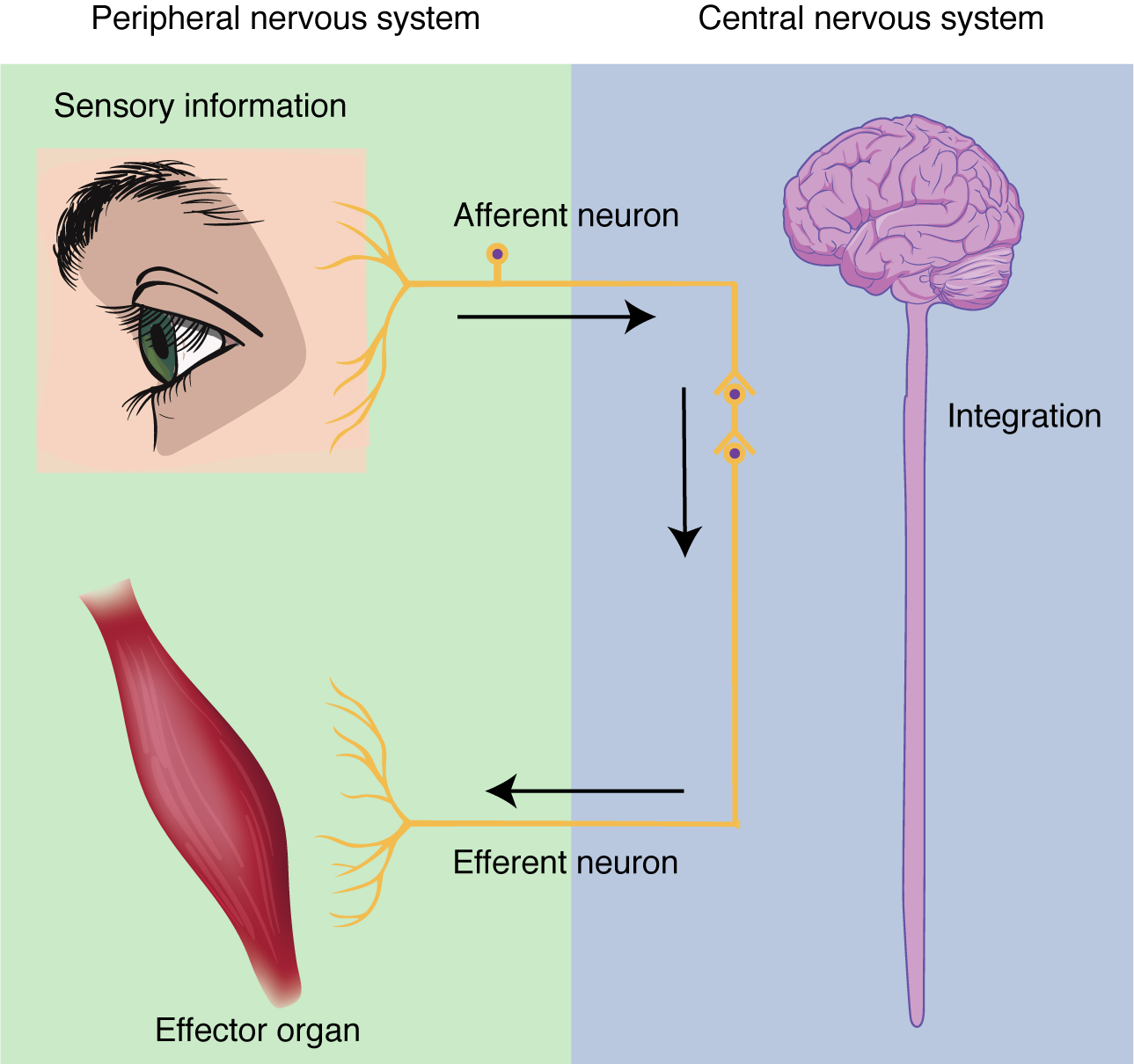
Sensation . Sensation refers to receiving information about the environment, either what is happening outside (ie: heat from the sun) or inside the body (ie: heat from muscle activity). These sensations are known as stimuli (singular = stimulus ) and different sensory receptors are responsible for detecting different stimuli. Sensory information travels towards the CNS through the PNS nerves in the specific division known as the afferent (sensory) branch of the PNS. When information arises from sensory receptors in the skin, skeletal muscles, or joints, it is transmitted to the CNS using somatic sensory neurons; when information arises from sensory receptors in the blood vessels or internal organs, it is transmitted to the CNS using visceral sensory neurons.
Response. The nervous system produces a response in effector organs (such as muscles or glands) due to the sensory stimuli. The motor ( efferent ) branch of the PNS carries signals away from the CNS to the effector organs. When the effector organ is a skeletal muscle, the neuron carrying the information is called a somatic motor neuron; when the effector organ is cardiac or smooth muscle or glandular tissue, the neuron carrying the information is called an autonomic motor neuron. Voluntary responses are governed by somatic motor neurons and involuntary responses are governed by the autonomic motor neurons, which are discussed in the next section.
Integration . Stimuli that are detected by sensory structures are communicated to the nervous system where information is processed. In the CNS, information from some stimuli is compared with, or integrated with, information from other stimuli or memories of previous stimuli. Then, a motor neuron is activated to initiate a response from the effector organ. This process during which sensory information is processed and a motor response generated is called integration (see Figure 12.1.2 below).
Chapter Review
The nervous system can be separated into divisions on the basis of anatomy and physiology. The anatomical divisions are the central and peripheral nervous systems. The CNS is the brain and spinal cord. The PNS is everything else and includes afferent and efferent branches with further subdivisions for somatic, visceral and autonomic function. Functionally, the nervous system can be divided into those regions that are responsible for sensation, those that are responsible for integration, and those that are responsible for generating responses.
Review Questions
Critical thinking questions.
1. What responses are generated by the nervous system when you run on a treadmill? Include an example of each type of tissue that is under nervous system control.
2. When eating food, what anatomical and functional divisions of the nervous system are involved in the perceptual experience?
Answers for Critical Thinking Questions
- Running on a treadmill involves contraction of the skeletal muscles in the legs (efferent somatic motor), increase in contraction of the cardiac muscle of the heart (efferent autonomic motor), and the production and secretion of sweat in the skin to stay cool (sensation of temp = afferent visceral sensory, sweat gland activation = efferent autonomic motor).
- The perceptual experience of eating food refers to tasting food, both in terms of flavors and texture. The neurons responsible for sensing taste are afferent somatic neurons of the PNS.
This work, Anatomy & Physiology, is adapted from Anatomy & Physiology by OpenStax , licensed under CC BY . This edition, with revised content and artwork, is licensed under CC BY-SA except where otherwise noted.
Images, from Anatomy & Physiology by OpenStax , are licensed under CC BY except where otherwise noted.
Access the original for free at https://openstax.org/books/anatomy-and-physiology/pages/1-introduction .
Anatomy & Physiology Copyright © 2019 by Lindsay M. Biga, Staci Bronson, Sierra Dawson, Amy Harwell, Robin Hopkins, Joel Kaufmann, Mike LeMaster, Philip Matern, Katie Morrison-Graham, Kristen Oja, Devon Quick, Jon Runyeon, OSU OERU, and OpenStax is licensed under a Creative Commons Attribution-ShareAlike 4.0 International License , except where otherwise noted.
Section 5: The Nervous System
Introduction to the nervous system, learning objectives.
- Identify the anatomical and functional organization of the nervous system
- Describe the functional and structural differences between gray matter and white matter structures
- Describe the basic structure of a neuron
- Identify the different types of neurons on the basis of polarity
- Identify the different functional types of neurons
- List the glial cells of the CNS and describe their function
- List the glial cells of the PNS and describe their function
- Distinguish the major functions of the nervous system: sensation, integration, and response
- Describe the structures at a synapse.
- Categorize the major neurotransmitters by chemical type and effect

Figure 1. Human Nervous System. The ability to balance like an acrobat combines functions throughout the nervous system. The central and peripheral divisions coordinate control of the body using the senses of balance, body position, and touch on the soles of the feet. (credit: Rhett Sutphin)
The nervous system is responsible for controlling much of the body, both through somatic (voluntary) and autonomic (involuntary) functions. The structures of the nervous system must be described in detail to understand how many of these functions are possible. There is a physiological concept known as localization of function that states that certain structures are specifically responsible for prescribed functions. It is an underlying concept in all of anatomy and physiology, but the nervous system illustrates the concept very well.
Fresh, unstained nervous tissue can be described as gray or white matter, and within those two types of tissue it can be very hard to see any detail. However, as specific regions and structures have been described, they were related to specific functions. Understanding these structures and the functions they perform requires a detailed description of the anatomy of the nervous system, delving deep into what the central and peripheral structures are.
The place to start this study of the nervous system is the beginning of the individual human life, within the womb. The embryonic development of the nervous system allows for a simple framework on which progressively more complicated structures can be built. With this framework in place, a thorough investigation of the nervous system is possible.
The nervous system is often referred to as the master controller of the human body. Like the endocrine system, the other internal control system of the human body, the nervous system is specialized for communication of information from one part of the body to another. The nervous system communicates quickly using neurons, the specialized cells of the nervous system. Neurons can convey and process information using electrical and chemical signals. Ultimately, neural communication helps coordinate body activities and ensures we maintain homeostasis.
The Components of the Nervous System
Imagine that you decide to bake a cake for your Anatomy and Physiology professor’s birthday. You find a recipe, locate the ingredients and then one by one, measure and add each to the mixing bowl. You stir the mixture and before you know it, the cake is in the oven and you can return to your homework. You start to salivate as a yummy chocolate aroma wafts through the room. The timer goes off, but unfortunately, when removing the pan from the oven you burn your hand through a hole in the hot pad. Rather than dropping the cake, you make a split second decision to hold on, enduring the pain so that you can set the cake pan down safely even though the consequences of a burnt hand will be with you for a few days. All aspects of baking a cake from your decision to bake a cake, to reading the recipe, measuring and mixing ingredients, smelling a delicious chocolate aroma and automatically starting to salivate in case you decide to sneak a piece of cake, and withdrawing your hand to prevent a burn are functions of the nervous system. Although quite different behaviors, they all include the general functions of the nervous system listed below:
- The nervous system detects changes in our internal and external environment (stimuli) using specific neurons or specialized cells communicating with neurons called sensory receptors.Sensory receptors can detect a variety of different external and internal stimuli such as: skin temperature, light (for vision), sound, chemicals in food (taste) and air (smells), pressure, pain, blood pH, core body temperature, bladder distension as well as many other stimuli.
- Sensory receptors transform stimuli into electric signals that our nervous system can understand. The nervous system cannot directly interpret stimuli like light, heat or sound. The information has to be transformed into an electrical signal for the nervous system to receive and process the information.
- Sensory neurons transmit the electrical signals from the periphery to the central nervous system (brain and spinal cord). This information travels from the sensory receptor (the site of transformation) along neural processes called axons towards the central nervous system (CNS).
- The central nervous system (brain and spinal cord) processes incoming sensory information to generate “appropriate” responses and also to give us the perception of the stimulus. The signal can be compared to a normal value (set point) or related to past experiences to determine if the stimulus requires a response. Processing of the signal is called integration. In order to perceive a stimulus, the sensory information must be transmitted to specific areas of the brain.
- The central nervous system sends commands (electrical signals passed along neurons) out to the target tissues to produce the response. The target tissues of the nervous system are muscles and glands.
- Anatomy & Physiology. Authored by : OpenStax College. Provided by : Rice University. Located at : http://cnx.org/contents/[email protected] . License : CC BY: Attribution . License Terms : Download for free at http://cnx.org/contents/[email protected]

- school Campus Bookshelves
- menu_book Bookshelves
- perm_media Learning Objects
- login Login
- how_to_reg Request Instructor Account
- hub Instructor Commons
- Download Page (PDF)
- Download Full Book (PDF)
- Periodic Table
- Physics Constants
- Scientific Calculator
- Reference & Cite
- Tools expand_more
- Readability
selected template will load here
This action is not available.
9.2: Introduction to the Nervous System
- Last updated
- Save as PDF
- Page ID 64638

- Suzanne Wakim & Mandeep Grewal
- Butte College
In the Blink of an Eye
As you drive into a parking lot, a skateboarder suddenly flies in front of your car across your field of vision. You see the skateboarder in the nick of time and react immediately. You slam on the brakes and steer sharply to the right — all in the blink of an eye. You avoid a collision, but just barely. You’re shaken up but thankful that no one was hurt. How did you respond so quickly? Such rapid responses are controlled by your nervous system.

Overview of the Nervous System
The nervous system , illustrated in Figure \(\PageIndex{2}\), is the human organ system that coordinates all of the body’s voluntary and involuntary actions by transmitting electrical signals to and from different parts of the body. Specifically, the nervous system extracts information from the internal and external environments using sensory receptors. It then usually sends signals encoding this information to the brain, which processes the information to determine an appropriate response. Finally, the brain sends signals to muscles, organs, or glands to bring about the response. In the example above, your eyes detected the skateboarder, the information traveled to your brain, and your brain instructed your body to act so as to avoid a collision.
Signals of the Nervous System
The signals sent by the nervous system are electrical signals called nerve impulses, and they are transmitted by special nervous system cells named neurons, or nerve cells, like the one in Figure \(\PageIndex{3}\) (all the parts of a neuron are explained in the next section). Dendrites of a neuron receive nerve impulses from other cells. Long projection (called axons) from neurons carries nerve impulses directly to specific target cells. Schwann cells wrapped around the axon are called glial cells . They create a myelin sheath which allows the nerve impulse to travel very rapidly through the axons. A cell that receives nerve impulses from a neuron may be excited to perform a function, inhibited from carrying out an action, or otherwise controlled. In this way, the information transmitted by the nervous system is specific to particular cells and is transmitted very rapidly.
In fact, the fastest nerve impulses travel at speeds greater than 100 meters per second! Compare this to the chemical messages carried by the hormones that are secreted into the blood by endocrine glands. These hormonal messages are “broadcast” to all the cells of the body, and they can travel only as quickly as the blood flows through the cardiovascular system.

Organization of the Nervous System
As you might predict, the human nervous system is very complex. It has multiple divisions, beginning with its two main parts, the central nervous system (CNS) and the peripheral nervous system (PNS), as shown in Figure \(\PageIndex{4}\). The CNS includes the brain and spinal cord, and the PNS consists mainly of nerves, which are bundles of axons from neurons. The nerves of the PNS connect the CNS to the rest of the body. You can learn much more about the CNS by reading the concept Central Nervous System .

The PNS is divided into two major parts, called the autonomic and somatic nervous systems. The somatic nervous system controls activities that are under voluntary control, such as turning a steering wheel. The autonomic nervous system controls activities that are not under voluntary control, such as digesting a meal. The autonomic nervous system has two divisions: the sympathetic division, which controls the fight-or-flight response during emergencies, and the parasympathetic division, which controls the routine “housekeeping” functions of the body at other times. You can learn more about the PNS and its subdivisions by reading the concept Peripheral Nervous System .
- List the general steps by which the nervous system generates an appropriate response to information from the internal and external environments.
- What are neurons?
- Compare and contrast the central and peripheral nervous systems.
- Which major division of the peripheral nervous system allows you to walk to class? Which major division of the peripheral nervous system controls your heart rate?
- Identify the functions of the three divisions of the autonomic nervous system.
- What is an axon and what is its function?
- True or False . A nerve impulse always causes the target cell to perform an action.
- True or False. The spinal cord is not considered part of the peripheral nervous system.
- Define nerve impulses.
- Explain why signals in the nervous system are generally more targeted and specific than signals in the endocrine system.
- Explain generally how the brain and spinal cord can interact with and control the rest of the body.
- ___________ actions are performed without the person thinking about them.
- autonomic nervous system
- somatic nervous system
- central nervous system
- parasympathetic nervous system
- How are nerves and neurons related?
- What type of information from the outside environment do you think is detected by sensory receptors in your ears?
Explore More
Attributions.
- Skateboarder by JESHOOTS-com via Pixabay license
- Nervous System diagram by the Emirr, CC BY 3.0 via Wikimedia Commons
- Neuron by NickGorton , licensed CC BY-SA 3.0 via Wikimedia Commons
- Nervous System Flowchart by Suzanne Wakim dedicated CC0
- Text adapted from Human Biology by CK-12 licensed CC BY-NC 3.0

- school Campus Bookshelves
- menu_book Bookshelves
- perm_media Learning Objects
- login Login
- how_to_reg Request Instructor Account
- hub Instructor Commons
- Download Page (PDF)
- Download Full Book (PDF)
- Periodic Table
- Physics Constants
- Scientific Calculator
- Reference & Cite
- Tools expand_more
- Readability
selected template will load here
This action is not available.
10.1: Introduction to the Nervous System
- Last updated
- Save as PDF
- Page ID 7174
Want to create or adapt books like this? Learn more about how Pressbooks supports open publishing practices.
56 Introduction to the Nervous System
Organization of the nervous system.
The nervous system is a network of cells called neurons that coordinate actions and transmit signals between different parts of the body.
Learning Objectives
Describe the organization of the nervous system
Key Takeaways
- Neurons (specialized cells of the nervous system ) send signals along thin fibers called axons and communicate with other cells by releasing chemicals called neurotransmitters at cell-cell junctions called synapses.
- Glial cells are non-neuronal cells that provide support and nutrition in the nervous system.
- In humans, the nervous system consists of both the central and peripheral nervous systems.
- The human central nervous system contains the brain, spinal cord, and retina.
- The peripheral nervous system consists of sensory neurons, clusters of neurons called ganglia, and nerves connecting them to each other and to the central nervous system.
- sensory receptor : A nerve ending that recognizes stimulus in the internal or external environment of an organism.
- peripheral nervous system : This system consists of the nerves and ganglia outside of the brain and spinal cord.
- glia : Non-neuronal cells that maintain homeostasis, form myelin, and provide support and protection for neurons in the brain and other parts of the nervous system.
A nervous system allows us to react to the changing environment around us.
The nervous system is an organ system that coordinates voluntary and involuntary actions and responses by transmitting signals between different parts of our bodies.
Central to the functioning of the nervous system is an extensive network of specialized cells called neurons. Neurons feature many thin projecting fibers called axons, which penetrate deep into tissues. They are able to communicate with other cells by chemical or electrical means at synapses. Neuronal function is supported by neuroglia, specialized cells which provide nutrition, mechanical support, and protection.

Major elements in neuron-to-neuron communication : Electrical impulses travel along the axon of a neuron. When this signal reaches a synapse, it provokes release of neurotransmitter molecules, which bind to receptor molecules located in the the target cell.
Divisions of the Nervous System
In most animals, including humans, the nervous system consists of two parts: central and peripheral. The central nervous system (CNS) is composed of the brain, spinal cord, and cerebellum. The peripheral nervous system (PNS) consists of sensory neurons, motor neurons, and neurons that communicate either between subdivisions of the PNS or connect the PNS to the CNS

The Human Nervous System : The major organs and nerves of the human nervous system. The CNS is comprised of the brain, cerebellum and spinal cord. Remaining neurons, and associated cells, distributed throughout the body form the PNS.
The nervous system has three broad functions: sensory input, information processing, and motor output. In the PNS, sensory receptor neurons respond to physical stimuli in our environment, like touch or temperature, and send signals that inform the CNS of the state of the body and the external environment. This sensory information is then processed by the CNS, predominantly by the brain.
After information is processed, motor neurons return signals to the muscles and glands of the PNS, which responds with motor output. Central neurons, which in humans greatly outnumber the sensory and motor neurons, make all of their input and output connections with other neurons. The connections of these neurons form neural circuits that are responsible for our perceptions of the world and determine our behavior. Along with neurons, the nervous system relies on the function of other specialized cells called glial cells, or glia, that provide structural and metabolic support to the nervous system.
Functions of the Nervous System
The primary function of the nervous system is to coordinate and control the various body functions.
Describe the functions of the nervous system
- The nervous system is a highly integrated system. The nervous system has three overlapping functions based on sensory input, integration, and motor output.
- At a more integrative level, the primary function of the nervous system is to control and communicate information throughout the body.
- hormone : A molecule released by a cell or a gland in one part of the body that sends out messages affecting cells in other parts of the organism.
- nervous system : The organ system that coordinates the activities of muscles, monitors organs, constructs and processes data received from the senses, and initiates actions.
The nervous system has three overlapping functions based on the sensory input, integration, and motor output. The nervous system is a highly integrated system.
Sensory Input
Sensory input comes from the many sensory receptors that monitor changes occurring both inside and outside the body. The total sum of the information gathered by these receptors is called sensory input. The nervous system processes and interprets sensory input and decides what actions should be taken. The nervous system activates effector organs such as muscles and glands to cause a response called motor output.
Integration
At a more integrative level, the primary function of the nervous system is to control and communicate information throughout the body. It does this by extracting information from the environment using sensory receptors. This sensory input is sent to the central nervous system, which determines an appropriate response.
Motor Response
Once the response is activated, the nervous system sends signals via motor output to muscles or glands to initiate the response.
In humans, the sophistication of the nervous system allows for language, abstract representation of concepts, transmission of culture, and many other features of society that would not otherwise exist.

Subdivisions of the Nervous System
The CNS includes the brain and spinal cord, while the PNS is a network of nerves linking the body to the brain and spinal cord.
Describe the subdivisions of the nervous system
- The nervous system is often divided into components called gray matter and white matter. Gray matter contains a relatively high proportion of neuron cell bodies and white matter is composed mainly of axons.
- The peripheral nervous system is subdivided into nerves, the autonomic system, and the somatic system. The autonomic nervous system is further subdivided into the parasympathetic and sympathetic nervous systems.
- The enteric nervous system is an independent subsystem of the peripheral nervous system.
- The central nervous system includes the brain and spinal cord and has various centers that integrate of all the information in the body. These can be subdivided into lower centers that carry out essential body functions and higher centers that control more sophisticated information processing.
- gray matter : A major component of the central nervous system, consisting of neuronal cell bodies, neuropil (dendrites and unmyelinated axons), glial cells (astroglia and oligodendrocytes), and capillaries.
- central nervous system : In vertebrates, the part of the nervous system comprising the brain, brainstem, and spinal cord.
- white matter : A region of the central nervous system containing myelinated nerve fibers and no dendrites.
The nervous system is comprised of two major subdivisions, the central nervous system (CNS) and the peripheral nervous system (PNS).
Central Nervous System
The Central Nervous System : The central nervous system (2) is a combination of the brain (1) and the spinal cord (3).
The CNS includes the brain and spinal cord along with various centers that integrate all the sensory and motor information in the body. These centers can be broadly subdivided into lower centers, including the spinal cord and brain stem, that carry out essential body and organ-control functions and higher centers within the brain that control more sophisticated information processing, including our thoughts and perceptions. Further subdivisions of the brain will be discussed in a later section.
Gray Matter and White Matter
The nervous system is often divided into components called gray matter and white matter. Gray matter, which is gray in preserved tissue but pink or light brown in living tissue, contains a relatively high proportion of neuron cell bodies. Conversely, white matter is composed mainly of axons and is named because of the color of the fatty insulation called myelin that coats many axons. White matter includes all of the nerves of the PNS and much of the interior of the brain and spinal cord. Gray matter is found in clusters of neurons in the brain and spinal cord and in cortical layers that line their surfaces.
By convention, a cluster of neuron cell bodies in the gray matter of the brain or spinal cord is called a nucleus, whereas a cluster of neuron cell bodies in the periphery is called a ganglion. However, there are a few notable exceptions to this rule, including a part of the brain called the basal ganglia, which will be discussed later.
Peripheral Nervous System
The PNS is a vast network of nerves consisting of bundles of axons that link the body to the brain and the spinal cord. Sensory nerves of the PNS contain sensory receptors that detect changes in the internal and external environment. This information is sent to the CNS via afferent sensory nerves. Following information processing in the CNS, signals are relayed back to the PNS by way of efferent peripheral nerves.
Autonomic and Somatic Nervous Systems
The PNS is further subdivided into the autonomic nervous system (ANS) and the somatic nervous system. The autonomic system has involuntary control of internal organs, blood vessels, and smooth and cardiac muscles. The somatic system has voluntary control of our movements via skeletal muscle.
As mentioned, the autonomic nervous system acts as a control system and most functions occur without conscious thought. The ANS affects heart rate, digestion, respiratory rate, salivation, perspiration, pupil diameter, urination, and sexual arousal. While most of its actions are involuntary, some, such as breathing, work in tandem with the conscious mind. The ANS is classically divided into two subsystems: the parasympathetic nervous system (PSNS) and sympathetic nervous system (SNS).
Parasympathetic and Sympathetic Nervous Systems
Broadly, the parasympathetic system is responsible for stimulation of “rest-and-digest” activities that occur when the body is at rest, including sexual arousal, salivation, lacrimation (tears), urination, digestion, and defecation. The sympathetic nervous syste is responsible for stimulating activities associated with the “fight-or-flight” response: mobilizing the systems of the body for escape or attacking sources of danger. In truth, the functions of both the parasympathetic and sympathetic nervous systems are not so straightforward, but this division is a useful rule of thumb.
The enteric nervous system (ENS) controls the gastrointestinal system and is sometimes considered part of the autonomic nervous system. However, it is sometimes considered an independent system because it can operate independently of the brain and the spinal cord.
The Nervous System of a Vertebrate : The brain and the spinal cord are the central nervous system (CNS) (shown in yellow). The left-right pair of cranial nerves, spinal nerves, and ganglia make up the peripheral nervous system (shown in dark gold).
Boundless Anatomy and Physiology Copyright © by Lumen Learning is licensed under a Creative Commons Attribution 4.0 International License , except where otherwise noted.
Share This Book
- Biology Article
- Nervous System
Human Nervous System
Living organisms adapt to their moves and positions in response to the environmental changes for their protection or to their advantage. When an entity reacts to the changes in its surroundings, it is referred to as stimulus while the reaction to the stimulus is referred to as a response. Common stimuli are sound, light, air, heat, smell, taste, water and gravity.
Think of burning your finger of fracturing your bone without any pain sensation. It may certainly sound like a superpower or an ideal situation, however, when it comes to the standpoint of survival, it can be disastrous.
The characteristic behaviour of living entities is to respond to stimuli with the intervention of the nervous system. It is an organ system ascribed to send signals from the spinal cord and the brain throughout the body and then back from all the body parts to the brain. The neuron acts as the mediator and is the basic signalling unit of the nervous system.
Pain is the body’s way of letting us know that something is not right. It can prevent further injuries or push us to seek medical attention. Moreover, all of this is possible because humans can respond and react to stimuli due to control and coordination among the various organs and organ systems.
Control and Coordination in simple multicellular organisms take place through only the Nervous system which coordinates activities of our body. It is the control system for all our actions, thinking, and behaviour.
Refer more: Control and Coordination
Let us have a detailed look at the nervous system notes to explore what is the nervous system, and the different functions of the nervous system with the help of diagrams. Table of Contents
What is the Nervous System?
Human nervous system diagram, central nervous system, peripheral nervous system, recommended video:.

The nervous system or the neural system is a complex network of neurons specialized to carry messages . The complexity of the nervous system increases as we move towards higher animals.
For instance, cnidarians such as jellyfish have relatively simple nerve nets spread throughout their body. Crabs have a more complicated nervous system in the form of 2 nerve centers called dorsal ganglion and ventral ganglion.
As we move further up the ladder, higher organisms such as vertebrates have a developed brain. Moreover, it is one of the most complicated structures in the animal kingdom, containing billions of neurons, all intricately connected.
In the human body, the neural system integrates the activities of organs based on the stimuli, which the neurons detect and transmit. They transmit messages in the form of electrical impulses and convey messages to and from the sense organs. Thus, the nervous coordination involves the participation of the sense organs, nerves, spinal cord, and brain.
Also Read: Creutzfeldt-Jakob Disease

Diagram of the Human Nervous System
One of the most complex organ system to ever evolve, the human nervous system consists of two parts, namely:
- Central Nervous System (consists of the brain and spinal cord)
- Peripheral Nervous System (includes all the nerves of the body)
Central Nervous System (CNS) is often called the central processing unit of the body. It consists of the brain and the spinal cord.
The brain is one of the important, largest and central organ of the human nervous system. It is the control unit of the nervous system, which helps us in discovering new things, remembering and understanding, making decisions, and a lot more. It is enclosed within the skull, which provides frontal, lateral and dorsal protection. The human brain is composed of three major parts:
Forebrain : The anterior part of the brain, consists of Cerebrum, Hypothalamus and Thalamus.
Midbrain : The smaller and central part of the brainstem, consists of Tectum and Tegmentum.
Hindbrain : The central region of the brain, composed of Cerebellum, Medulla and Pons.
Also read: Human Brain
Spinal Cord
The spinal cord is a cylindrical bundle of nerve fibers and associated tissues enclosed within the spine and connect all parts of the body to the brain. It begins in continuation with the medulla and extends downwards. It is enclosed in a bony cage called vertebral column and surrounded by membranes called meninges. The spinal cord is concerned with spinal reflex actions and the conduction of nerve impulses to and from the brain.
Peripheral Nervous System (PNS) is the lateral part of the nervous system that develops from the central nervous system which connects different parts of the body with the CNS. We carry out both voluntary and involuntary actions with the help of peripheral nerves.
Also refer: Peripheral Nervous System
PNS includes two types of nerve fibers:
- Afferent nerve fibers – These are responsible for transmitting messages from tissues and organs to the CNS.
- Efferent nerve-fibers – These are responsible for conveying messages from CNS to the corresponding peripheral organ.
Classification of the peripheral nervous system:
Somatic neural system (SNS): It is the neural system that controls the voluntary actions in the body by transmitting impulses from CNS to skeletal muscle cells. It consists of the somatic nerves.
Autonomic neural system (ANS): The autonomic neural system is involved in involuntary actions like regulation of physiological functions (digestion, respiration, salivation, etc.). It is a self-regulating system which conveys the impulses from the CNS to the smooth muscles and involuntary organs (heart, bladder and pupil). The autonomic neural system can be further divided into:
- Sympathetic nervous system
- Parasympathetic nervous system

A Neuron is a structured and functional unit of the nervous system and unlike other cells, neurons are irregular in shape and able to conduct electrochemical signals. The different parts of a neuron are discussed below.
- Dendrite stretches out from the cell body of a neuron, and it is the shortest fibre in the cell body.
- Axon is the longest thread on the cell body of a neuron and has an insulating and protective sheath of myelin around it.
- Cell body consists of cytoplasm and nucleus.
- Synapse is the microscopic gap between a pair of adjacent neurons over which nerve impulses pass, when moving from one neuron to the other.
Explore more: Placebo Effect
Nerves are thread-like structures that emerge from the brain and spinal cord. It is responsible for carrying messages to all the parts of the body. There are three types of nerves. Some of these neurons can fire signals at speeds of over 119 m/s or above 428 km/h.
- Sensory nerves send messages from all the senses to the brain.
- Motor nerves carry messages from the brain to all the muscles.
- Mixed nerves carry both sensory and motor nerves.
Also read: Nerves
Cranial nerves begin from the brain as these nerves carry impulses to start from the central nervous system. Certain cranial nerves belong to the group of mixed nerves while certain ones fall under sensory nerves. Spinal nerves originate from the spinal cord. All the spinal nerves carry impulses to and from the central nervous system and these are part of mixed nerves. The above nervous system diagram depicts the various nerves arising from various parts of the body.
Learn more in detail about the Human Nervous System with diagrams or any other related topics by referring to the nervous system notes provided at BYJU’S website. Download BYJU’S app for further reference.
Frequently Asked Questions
What are the two divisions of the nervous system.
The human nervous system controls all activities of the body in a quicker fashion. It can be divided into the central nervous system and peripheral nervous system. The central nervous system includes spinal cord and brain and the peripheral covers the nerves branching from spinal cord and brain.
What are nerves and neurons?
Nerves are thread-like structures that emerge from the spinal cord and brain. These nerves are actual projections of neurons. A neuron is a basic structural and functional unit of a nervous system that conducts electrochemical signals.
What are cranial nerves?
The nerves that extend throughout the body on both sides and emerges directly from the brain stem and brain are called cranial nerves. They carry information from the brain to other parts, primarily to the neck and head.

Put your understanding of this concept to test by answering a few MCQs. Click ‘Start Quiz’ to begin!
Select the correct answer and click on the “Finish” button Check your score and answers at the end of the quiz
Visit BYJU’S for all Biology related queries and study materials
Your result is as below
Request OTP on Voice Call
Leave a Comment Cancel reply
Your Mobile number and Email id will not be published. Required fields are marked *
Post My Comment
Hello Byju’s I need to chat with you to clarify a doubt. Your app is so good
Really Byjus is a very good application in the case of study in a systematic knowledge so Byjus achievement is great
Thank you for helping with this concept, it gave me better information than other websites.
THIS IS SERIOUSLY A NICE SITE FOR KNOWLEDGE.
Thanks a lot for helping me
Thanks a lot for helping
This app is very awesome and also help me in doing my homework ..
Actually I need more of these to pass my test thanks for sharing these notes I am really grateful to you about these Bye
Byju’s always help me a lot. .This platform is really great.
- Share Share
Register with BYJU'S & Download Free PDFs
Register with byju's & watch live videos.

IMAGES
VIDEO
COMMENTS
Nervous systems are of two general types, diffuse and centralized. In the diffuse type of system, found in lower invertebrates, there is no brain, and neurons are distributed throughout the organism in a netlike pattern.In the centralized systems of higher invertebrates and vertebrates, a portion of the nervous system has a dominant role in coordinating information and directing responses.
Introduction to the Nervous System. The nervous system is the major controlling, regulatory, and communicating system in the body. It is the center of all mental activity including thought, learning, and memory. Together with the endocrine system, the nervous system is responsible for regulating and maintaining homeostasis.Through its receptors, the nervous system keeps us in touch with our ...
The nervous system has two main parts: Central nervous system (CNS) : Your brain and spinal cord make up your CNS. Your brain reads signals from your nerves to regulate how you think, move and feel. Peripheral nervous system (PNS): Your PNS is made up of a network of nerves. The nerves branch out from your spinal cord.
It has multiple divisions, beginning with its two main parts, the central nervous system (CNS) and the peripheral nervous system (PNS), as shown in Figure 11.2.4 11.2. 4. The CNS includes the brain and spinal cord, and the PNS consists mainly of nerves, which are bundles of axons from neurons. The nerves of the PNS connect the CNS to the rest ...
The nervous system is a network of neurons whose main feature is to generate, modulate and transmit information between all the different parts of the human body. This property enables many important functions of the nervous system, such as regulation of vital body functions ( heartbeat, breathing, digestion), sensation and body movements.
Nervous tissue, present in both the CNS and PNS, contains two basic types of cells: neurons and glial cells. A glial cell is one of a variety of cells that provide a framework of tissue that supports the neurons and their activities. The neuron is the more functionally important of the two, in terms of the communicative function of the nervous system.
In biology, the nervous system is the highly complex part of an animal that coordinates its actions and sensory information by transmitting signals to and from different parts of its body. The nervous system detects environmental changes that impact the body, then works in tandem with the endocrine system to respond to such events. Nervous tissue first arose in wormlike organisms about 550 to ...
This page titled 11.1: Introduction to the Nervous System is shared under a CC BY license and was authored, remixed, and/or curated by Whitney Menefee, Julie Jenks, Chiara Mazzasette, & Kim-Leiloni Nguyen ( ASCCC Open Educational Resources Initiative) . The nervous system is a very complex organ system. The anatomy of the nervous system was ...
The nervous system is divided into two main parts: the central nervous system, consisting of the brain and spinal cord, and the peripheral nervous system, made up of nerves and ganglia. ... And I'll just mention some of the big ones for this kind of introduction talk. Here we're looking at the brain from the left side. All of this part on the ...
The nervous system is responsible for controlling much of the body, both through somatic (voluntary) and autonomic (involuntary) functions. The structures of the nervous system must be described in detail to understand how many of these functions are possible. There is a physiological concept known as localization of function that states that ...
The. nervous system. , illustrated in the sketch below, is the human organ system that coordinates all of the body's. voluntary. and. involuntary. actions, by transmitting electrical signals to and from different parts of the body. Specifically, the nervous system extracts information from the internal and external environments, using.
TRY IT FOR FREE TODAY: https://3d4med.com/331idBZDiscover the functions of the nervous system, and how it can be divided into the central nervous system and ...
The nervous system produces a response in effector organs (such as muscles or glands) due to the sensory stimuli. The motor ( efferent) branch of the PNS carries signals away from the CNS to the effector organs. When the effector organ is a skeletal muscle, the neuron carrying the information is called a somatic motor neuron; when the effector ...
The central nervous system (brain and spinal cord) processes incoming sensory information to generate "appropriate" responses and also to give us the perception of the stimulus. The signal can be compared to a normal value (set point) or related to past experiences to determine if the stimulus requires a response.
37 Nervous System Introduction In this unit, we'll explore the structure and function of the nervous system. The nervous system is often referred to as the master controller of the human body. Like the endocrine system, the other internal control system of the human body, the nervous system is specialized for communication of information from ...
This page titled 10.1: Introduction to the Nervous System is shared under a CC BY-NC-SA 4.0 license and was authored, remixed, and/or curated by Jennifer Lange et al.. The nervous system is a very complex organ system. The anatomy of the nervous system was first studied by Egyptians in the 300 BC.
The central nervous system processes information from various sources and responds to internal and external stimuli. It consists of the brain and spinal cord. Brain: The brain is the body's control center. It controls a myriad of body functions, from thoughts, reasoning, and language, to regulation of breathing and heart rate.
The nervous system, illustrated in Figure 9.2.2 9.2. 2, is the human organ system that coordinates all of the body's voluntary and involuntary actions by transmitting electrical signals to and from different parts of the body. Specifically, the nervous system extracts information from the internal and external environments using sensory ...
10.1: Introduction to the Nervous System - Medicine LibreTexts. search Search. build_circle Toolbar. fact_check Homework. cancel Exit Reader Mode. school Campus Bookshelves. menu_book Bookshelves. perm_media Learning Objects. login Login.
Nervous system 1: introduction to the nervous system | Nursing Times. Abstract This article, the first in a six-part series, provides an introduction to the nervous system. In particular, it explores the different cell types.
The central nervous system (CNS) is composed of the brain, spinal cord, and cerebellum. The peripheral nervous system (PNS) consists of sensory neurons, motor neurons, and neurons that communicate either between subdivisions of the PNS or connect the PNS to the CNS. The Human Nervous System: The major organs and nerves of the human nervous system.
In the human body, the neural system integrates the activities of organs based on the stimuli, which the neurons detect and transmit. They transmit messages in the form of electrical impulses and convey messages to and from the sense organs. Thus, the nervous coordination involves the participation of the sense organs, nerves, spinal cord, and ...
Introduction . Central nervous system (CNS)-related disorders are increasingly being recognized as a global health challenge worldwide. There are significant challenges for effective diagnosis and treatment due to the presence of the CNS barriers which impede the management of neurological diseases. Combination of nanovesicles (NVs) and ...